|
Case Report
Retinoblastoma in an 8-year-old boy: A rare case report
1 Department of Ophthalmology, King Fahad Hospital of the University, Al Khobar, Saudi Arabia
2 Department of Ophthalmology, Dhahran Eye Specialist Hospital, Dhahran, Saudi Arabia
3 College of Medicine, Princess Nourah bint Abdulrahman University, Riyadh, Saudi Arabia
4 College of Medicine, King Faisal University, Al-Ahsa, Saudi Arabia
Address correspondence to:
Nawaf AlJaafar
AlRakkah, AlKhobar 34225,
Saudi Arabia
Message to Corresponding Author
Article ID: 100054Z17RA2026
Access full text article on other devices

Access PDF of article on other devices


How to cite this article
AlAkel R, AlJaafar N, AlShaibani A, AlNfaiei J, AlKhalifa F, Emara K. Retinoblastoma in an 8-year-old boy: A rare case report. J Case Rep Images Opthalmol 2026;9(2):1–5.ABSTRACT
Retinoblastoma is a rare intraocular malignant tumor, most commonly seen in children under five years of age. We report a rare case of retinoblastoma in an 8-year-old boy who experienced right eye pain and blurring of vision for one month. The right eye visual acuity was 20/38. Slit-lamp examination revealed conjunctival injection, with floating nodules at the periphery of the anterior chamber and pseudohypopyon. The remainder of the examination was otherwise unremarkable. B-scan of the affected eye showed dense vitreous opacity and an inferonasal retinal mass with underlying exudative retinal detachment. An exploratory Pars Plana Vitrectomy (PPV) with vitreous sampling was performed to rule out intraocular lymphoma, and histopathological assessment revealed malignant cells, confirming the diagnosis of retinoblastoma. The patient subsequently underwent immediate enucleation after obtaining informed consent from his family. Following enucleation, the patient is doing well with a prosthetic eye. To the best of our knowledge, this is the first reported case of an atypical presentation of retinoblastoma in the Saudi population.
Keywords: Children, Late onset, Leukocoria, Retinoblastoma
Introduction
Retinoblastoma (RB) is the most common primary intraocular malignancy in children, resulting from immature retinal cells. It is reported to affect one in 15,000 to 20,000 live births [1],[2]. Retinoblastoma can be heritable, frequently due to germline mutations of RB1, or sporadic, secondary to MYCN amplification [2]. It often comes to clinical attention when patients develop leukocoria; however, a subset of patients may present with complaints of squint, redness, swelling, or visual scintillations [2]. Approximately 95% of cases are identified in the first five years of life, and diagnosis at an age greater than five years is rare and difficult. Unusual late manifestations may result in delayed diagnosis and considerably worse outcomes, including parental stress, local invasion, higher rates of enucleation, and distant metastasis [1],[3]. Multimodal treatments such as systemic chemotherapy, intra-arterial chemotherapy, and local treatments have made tremendous progress in recent times, focusing on achieving survival rate improvement and visual preservation [3]. In this paper, we present an unusual case of retinoblastoma in an 8-year-old boy, as this malignancy typically presents in much younger children.
Case Report
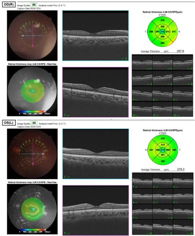
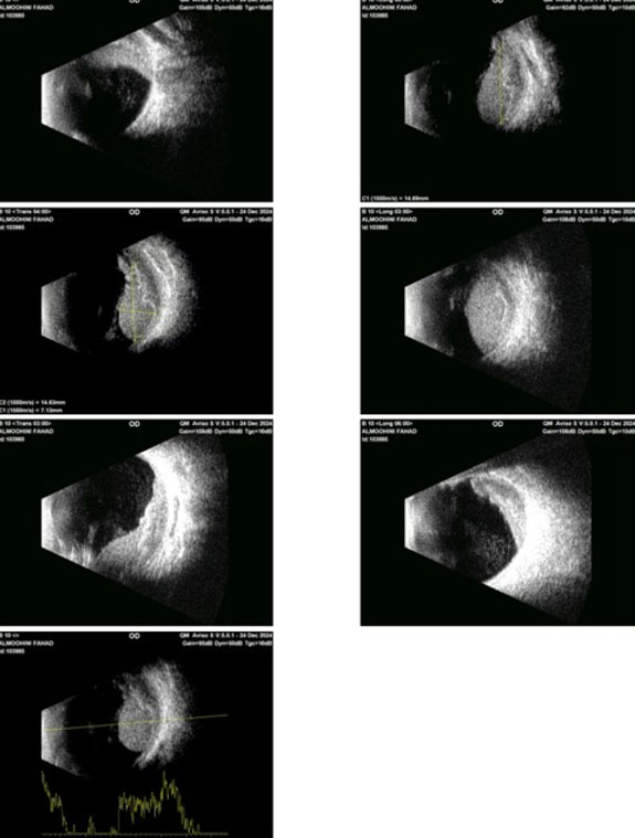
On 24 December 2024, an 8-year-old boy, medically and surgically free, presented to Dhahran Eye Specialist Hospital (DESH) with a history of right eye pain and blurring of vision over the past one month, and was referred to the uveitis clinic for further evaluation. The onset of symptoms was reported after swimming in a pool. The patient denied any previous similar episodes. He is not on any medication and has no known allergies. On examination, the visual acuity of the right eye was 20/38, and the left eye was 20/20. The slit-lamp examination of the right eye revealed a mild conjunctival injection, anterior chamber is deep with white floating nodules resting in the periphery of the angle and there is pseudohypopyon, the cornea was clear with no keratic precipitates, and the lens was clear as well. The left eye is unremarkable with no significant finding. Fundus photo of the right eye was done and showed a large nasal white vitreous mass occupying nearly the whole nasal retina. Optical coherence tomography (OCT) macula was within the normal limit for both eyes (Figure 1). However, since the OCT was only coveting the posterior pole macular region, it was not able to locate this lesion since it was far on the nasal side. To complete the assessment, a B-scan was done and revealed mildly dense vitreous opacities and a retinal mass located in the inferonasal area measuring 14.63 mm wide, 14.69 mm long, and 7.13 mm high. There was exudative retinal detachment (ERD) beneath the mass, not involving the macula. The optic nerve head (ONH) appeared normal (Figure 2). The patient was referred to a pediatric ophthalmology clinic, where they suspected fungal endophthalmitis vs intraocular lymphoma. Since the diagnosis was not clear, examination under anesthesia (EUA) for both eyes (oculus uterque, OU) and anterior chamber (AC) sampling of aqueous humor were decided for this patient and performed on the 26th of December successfully with no complications. Conservative treatment was attempted with intravitreal methotrexate in the right eye (oculus dexter, OD) under general anesthesia. There were no complications, and the patient was discharged.
On December 29th, a B-scan ultrasound showed no increase in the mass’s size. On January 7th, the patient was planned for an exploratory pars plana vitrectomy (PPV) and vitreous sampling in the right eye (OD) by the surgical retina team. The surgery was performed on January 9th. Postoperatively, the patient had visual acuity (VA) of hand motion (HM), intraocular pressure (IOP) of 20 mmHg, +++ cells in the anterior chamber (AC), and the fundus could not be visualized due to the presence of air. On January 13th, histopathological examination revealed malignant cells, likely indicating retinoblastoma. The patient was sent to King Fahad Specialist Hospital for complete oncology work-up and possible initiation of chemotherapy treatment.
After that, surgery was indicated to reduce the risk of local extension and distant metastases without needing chemotherapy.
On January 16th, enucleation was performed by the oculoplastic team. No alternative surgical options were considered. The risk-benefit discussion concluded that immediate enucleation was necessary to minimize the risk of spread and save the patient’s life. There were no preoperative or intraoperative challenges, and no complications were reported.
Surgical Pathology Report (performed at King Fahad Specialist Hospital):
– Right Eye Enucleation. Diagnosis: Retinoblastoma, poorly differentiated (Grade 4). Pathologic Findings: Tumor Size: 1.4 cm in maximum dimension. Iris Stroma: Involved by tumor. Optic Nerve (including lamina cribrosa): Free of tumor involvement. Pathologic Staging (AJCC 8th Edition – pTNM): Primary Tumor: pT2b Regional Lymph Nodes: not assessed Distant Metastasis: not assessed.
The patient recovered well postoperatively and was discharged on January 19th. Follow-up on January 23rd showed the patient doing well with the conformer in place and advised to use protective glasses and to have regular follow-ups with anaplastologists.
Discussion
Retinoblastoma, while the most common primary intraocular cancer in children, is still considered a rare pediatric tumor. Typically, bilateral cases are diagnosed before one year of age and unilateral cases before two years [4]. When the disease appears in older children, it is much more likely to be misdiagnosed. This happens not only because doctors are less likely to suspect retinoblastoma in this age group, but also because older children often show unusual symptoms. These may include hyphema, pseudohypopyon, vitreous bleeding, staphyloma, new abnormal blood vessels on the iris, cataract, glaucoma, or even a shrinking eye—features that are rarely seen in typical retinoblastoma [5]. A retrospective observational study was conducted at New Delhi, India, in which 48 out of 610 (7.8%) patients with retinoblastoma were older than 6 years. 22/48 (46%) had atypical symptoms including proptosis, glaucoma, neovascularization of the iris, and staphyloma as the most frequent presentations. 14/48 (29%) cases were misdiagnosed initially and the most common misdiagnosis was endophthalmitis. Initial misdiagnosis in older age retinoblastoma may result in a delayed referral and consequent advanced disease at presentation [3]. Moreover, a case reported on June, 2018 from Hospital Kuala Lumpur, Kuala Lumpur, Malaysia, had discussed the unique presentation of a 9-year-old girl who had sudden loss of vision of the left eye following a fall at home. Examination of the left eye revealed an extensive ERD that was confirmed by B-scan. Afterward, computed tomography (CT) scan was done and showed a left eye intraocular mass with calcification. Examination under anesthesia revealed a mixed endophytic and exophytic mass with extensive exudative retinal detachment. Family members consented to left eye enucleation and histopathological report confirmed the diagnosis of retinoblastoma [6]. Our case is one of the first case reports highlighting an atypical late childhood presentation of retinoblastoma. It is relevant due to the relative rareness of this disease in this age group, which will allow clinicians to think about this disease as a possible, albeit rare, diagnosis possibility. The patient’s presentation with eye pain and decreased vision brings into account the importance of applying a full ophthalmic examination and having a high clinical suspicion. Our case herein brings into attention the importance of considering retinoblastoma in the differential diagnosis of any unclear intra-ocular pathology in children. When there is uncertainty in diagnosis, the clinicians need to weight the benefits of enucleation against the risks without enucleation. Certainly, enucleation of one eye will cause significant psychological and social impact on the affected children. However, failure to diagnose and treat retinoblastoma may lead to significant morbidity and even mortality to the patients. It is still safer to offer enucleation when there is diagnosis uncertainty and retinoblastoma is suspected [4]. From this case, several lessons emerge. First, in older pediatric Rb patients, symptoms and signs are often atypical for Rb, such as conjunctival edema, a pseudohypopyon in the anterior chamber, pseudocellulitis of the orbit, and secondary intraocular glaucoma. Patients with these symptoms are easily misdiagnosed as uveitis or infectious endophthalmitis. However, the pseudohypopyon seen in retinoblastoma differs from the thick yellow-white hypopyon of infectious endophthalmitis; instead, it consists of gray-white, granular, or sponge-like tumor cells that settle in the lower part of the anterior chamber or diffusely seeded in the anterior chamber, the surface of the iris or the surface of the lens. Second, in suspected intraocular malignancy with atypical findings and inconclusive imaging, anterior chamber or vitreous fluid analysis should be performed promptly to secure the diagnosis. Finally, the primary goal of retinoblastoma management is to save the patient’s life, followed by preservation of the eye and visual function.
Conclusion
The primary teaching point of this case report is to keep in mind that retinoblastoma presentation could be atypical, and that treating clinician must maintain a high index of suspicion for retinoblastoma in older children in order to avoid misdiagnosis, inadvertent surgery, and mortality from this disease entity. This case may alter clinical practice in the future to include it within the differential diagnosis of older children with unexplained visual changes.
REFERENCES
1.
Rao R, Honavar SG. Retinoblastoma. Indian J Pediatr 2017;84(12):937–44. [CrossRef]
[Pubmed]

2.
Atima-Ayeni E, Orugun AJ, Idakwo U, Komolafe O, Atima MO, Atanda AT, et al. Retinoblastoma in a young Nigerian girl: A case report from ECWA Eye Hospital, Kano. Case Rep Ophthalmol Med 2025;2025:1733830. [CrossRef]
[Pubmed]
3.
Meel R, Kashyap S, Bakhshi S, Singh Bajaj M, Wadhwani M. Retinoblastoma in children older than 6 years of age. Ocul Oncol Pathol 2020;6(6):395–404. [CrossRef]
[Pubmed]
4.
Cassoux N, Lumbroso L, Levy-Gabriel C, Aerts I, Doz F, Desjardins L. Retinoblastoma: Update on current management. Asia Pac J Ophthalmol (Phila) 2017;6(3):290–8. [CrossRef]
[Pubmed]
5.
Karcioglu ZA, Abboud EB, Al-Mesfer SA, Al-Rashed W, Pilapil DH. Retinoblastoma in older children. J AAPOS 2002;6(1):26–32. [CrossRef]
[Pubmed]
6.
Ong WZ, Rahmat J, Ramasamy S, Samsudin A. Retinoblastoma in an older child: A case report. Mal J Med Health Sci 2018;14(2):93–96.
SUPPORTING INFORMATION
Acknowledgments
We would like to extend our gratitude to the Dhahran Eye Specialist Hospital, Dhahran, Saudi Arabia, for the macular optical coherence tomography and the ultrasound biometry.
Author ContributionsRaghad AlAkel - Conception of the work, Design of the work, Revising the work critically for important intellectual content, Final approval of the version to be published, Agree to be accountable for all aspects of the work in ensuring that questions related to the accuracy or integrity of any part of the work are appropriately investigated and resolved.
Nawaf AlJaafar - Drafting the work, Final approval of the version to be published, Agree to be accountable for all aspects of the work in ensuring that questions related to the accuracy or integrity of any part of the work are appropriately investigated and resolved.
Askar AlShaibani - Revising the work critically for important intellectual content, Final approval of the version to be published, Agree to be accountable for all aspects of the work in ensuring that questions related to the accuracy or integrity of any part of the work are appropriately investigated and resolved.
Judi AlNfaiei - Acquisition of data, Drafting the work, Final approval of the version to be published, Agree to be accountable for all aspects of the work in ensuring that questions related to the accuracy or integrity of any part of the work are appropriately investigated and resolved.
Fatemah AlKhalifa - Analysis of data, Drafting the work, Final approval of the version to be published, Agree to be accountable for all aspects of the work in ensuring that questions related to the accuracy or integrity of any part of the work are appropriately investigated and resolved.
Khalid Emara - Conception of the work, Design of the work, Revising the work critically for important intellectual content, Final approval of the version to be published, Agree to be accountable for all aspects of the work in ensuring that questions related to the accuracy or integrity of any part of the work are appropriately investigated and resolved.
Guaranter of SubmissionThe corresponding author is the guarantor of submission.
Source of SupportNone
Consent StatementWritten informed consent was obtained from the patient for publication of this article.
Data AvailabilityAll relevant data are within the paper and its Supporting Information files.
Conflict of InterestAuthors declare no conflict of interest.
Copyright© 2026 Raghad AlAkel et al. This article is distributed under the terms of Creative Commons Attribution License which permits unrestricted use, distribution and reproduction in any medium provided the original author(s) and original publisher are properly credited. Please see the copyright policy on the journal website for more information.