|
Case Report
Delayed-onset iris prolapse 1-month following uncomplicated cataract surgery
1 BS, Medical Student, The University of Texas at Tyler School of Medicine, Tyler, Texas, USA
2 MD, Volunteer Community Faculty, The University of Texas at Tyler School of Medicine; Ophthalmologist, Heaton Eye Associates, Tyler, Texas, USA
3 OD, Optometrist, Heaton Eye Associates, Tyler, Texas, USA
Address correspondence to:
Shrina Patel
3355 Earl Campbell Parkway, Tyler, TX 75701,
USA
Message to Corresponding Author
Article ID: 100058Z17SP2026
Access full text article on other devices

Access PDF of article on other devices

How to cite this article
Patel S, Dossey A, Marshall M. Delayed-onset iris prolapse 1-month following uncomplicated cataract surgery. J Case Rep Images Opthalmol 2026;9(2):23–27.ABSTRACT
Introduction: Iris prolapse is a well-recognized complication of cataract surgery that typically occurs intraoperatively or in the immediate operative period. It results from a combination of predisposing risk factors, surgical technique and instruments, pharmacologic use, and patient-related behavioral factors such as eyelid squeezing and poor compliance with postoperative activity restrictions.
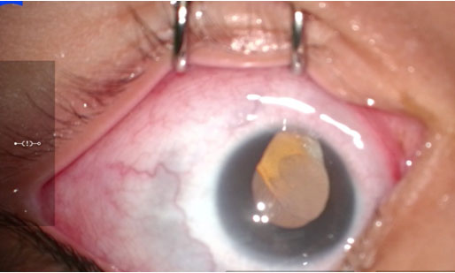
Case Report: A 69-year-old female presented to the clinic for a one-month postoperative follow-up after uncomplicated cataract surgery of the right eye (OD). The patient was subjectively asymptomatic but had noticed a “dark spot” on her globe, which was initially mistaken for cosmetic debris. Slit-lamp examination revealed a prolapse of iris tissue through the temporal corneal incision with associated peaked pupil. The patient returned to the operating room the next morning where the iris was repositioned, followed by intracameral air bubble insertion and placement of a temporal suture. Surgery was uncomplicated and visual acuity the following day had returned to 20/20.
Conclusion: Iris prolapse can present as a rare, asymptomatic late complication of cataract surgery. This case highlights the significance of longitudinal wound assessment and patient education regarding accidental ocular trauma, even after a seemingly stable early postoperative course. Urgent surgical repair is necessary to prevent further complications, such as permanent iris necrosis, chronic pupillary distortion, or the development of endophthalmitis.
Keywords: Cataract surgery, Clear corneal incision, Delayed- onset, Iris prolapse
SUPPORTING INFORMATION
Acknowledgments
Artificial Intelligence (AI) Disclosure:
Generative AI technology (Google Gemini, May 2026 version, Google LLC) was utilized during the preparation of this manuscript. Specifically, the technology was used to assist with grammatical editing and formatting of the literature citations within the manuscript text. The final clinical interpretation and critical content verification were performed entirely by the authors, who accept full accountability for the integrity and accuracy of the content.
Shrina Patel - Substantial contributions to conception and design, Acquisition of data, Analysis of data, Interpretation of data, Drafting the article, Revising it critically for important intellectual content, Final approval of the version to be published
Adam Dossey - Substantial contributions to conception and design, Acquisition of data, Analysis of data, Interpretation of data, Drafting the article, Revising it critically for important intellectual content, Final approval of the version to be published
Matthew Marshall - Substantial contributions to conception and design, Acquisition of data, Analysis of data, Interpretation of data, Drafting the article, Revising it critically for important intellectual content, Final approval of the version to be published
Guaranter of SubmissionThe corresponding author is the guarantor of submission.
Source of SupportNone
Consent StatementWritten informed consent was obtained from the patient for publication of this article.
Data AvailabilityAll relevant data are within the paper and its Supporting Information files.
Conflict of InterestAuthors declare no conflict of interest.
Copyright© 2026 Shrina Patel et al. This article is distributed under the terms of Creative Commons Attribution License which permits unrestricted use, distribution and reproduction in any medium provided the original author(s) and original publisher are properly credited. Please see the copyright policy on the journal website for more information.