|
Case Report
Ophthalmic manifestations of MAB21L2 mutation: A case report
1 Al Bahar Eye Center, Ibn Sina Hospital, Ministry of Health, Al Shuwaikh, Kuwait City, Kuwait
2 Residency Program Director, Kuwait Board of Ophthalmology, Kuwait Institute for Medical Specialization, Jamal Abdulnaser Street, Sulibekhat, Kuwait City, Kuwait
3 Dasman Diabetes Institute, Gulf Road intersecting Jassim Al Bahar Street, Sharq, Kuwait City, Kuwait
Address correspondence to:
AbdalWahab AlEnezy
Al Bahar Eye Center, Ibn Sina Hospital, Ministry of Health, Al Shuwaikh, Kuwait City,
Kuwait
Message to Corresponding Author
Article ID: 100056Z17AA2026
Access full text article on other devices

Access PDF of article on other devices

How to cite this article
AlEnezy A, AlKandari A, AlAli A. Ophthalmic manifestations of MAB21L2 mutation: A case report. J Case Rep Images Opthalmol 2026;9(2):11–16.ABSTRACT
Introduction: This report is to describe the clinical presentation, ophthalmic findings, and multimodal evaluation of a child with severe bilateral microphthalmia and coloboma associated with a MAB21L2 variant.
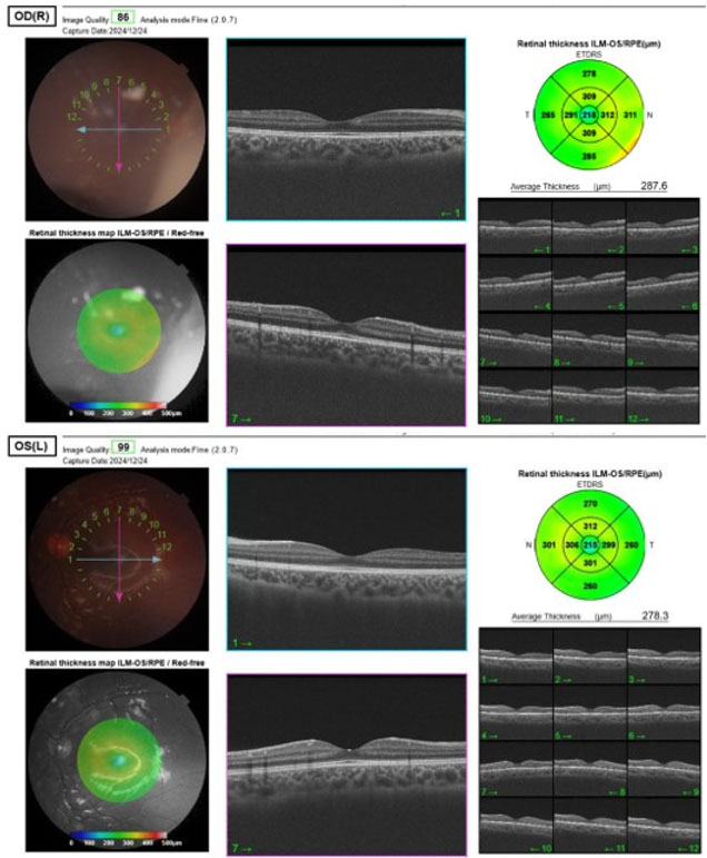
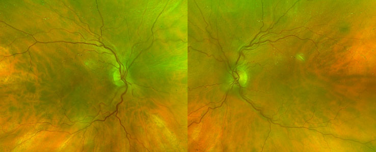
Case Report: A 3-year-old girl presented with bilateral small eyes and abnormal eye movements. Examination showed chin-up posture, bilateral ptosis, large-angle non-accommodative esotropia, limitation of abduction, wandering nystagmus, xanthocoria on Bruckner test, microcornea [8 mm OD (right eye), 6.5 mm OS (left eye)), sclerocornea superiorly with clear central cornea, bilateral inferonasal iris coloboma, and inferior lens subluxation with zonular disruption. Fundus examination revealed a severely disorganized retina with retinal cysts and folds and inferior chorioretinal coloboma. Intraocular pressure (IOP) was 21 mmHg OD and 19 mmHg OS. A B-scan revealed taut retinal folds with a retinal cyst in the OD and a retinal cyst in the OS. Magnetic resonance imaging (MRI) orbit/brain showed small globes [measuring about 11–12 mm in anteroposterior diameter (AP)], with multiple retrobulbar cysts encroaching on the optic nerve sheath complex, bilateral optic nerve hypoplasia (T p.(Cys250Phe).
Conclusion: This case illustrates a severe microphthalmia and coloboma spectrum disorder with extensive anterior and posterior segment dysgenesis and marked optic nerve hypoplasia. Serial examination under anesthesia (EUA) and multimodal imaging were of the essence for complete characterization and follow-up. The familial pattern and MAB21L2 finding provide a unifying developmental explanation for the case.
Keywords: Coloboma, MAB21L2 mutation, Microphthalmia, Ophthalmic manifestations, Optic nerve hypoplasia
SUPPORTING INFORMATION
Author Contributions
AbdalWahab AlEnezy - Substantial contributions to conception and design, Acquisition of data, Interpretation of data, Drafting the article, Revising it critically for important intellectual content, Final approval of the version to be published
Aseel AlKandari - Acquisition of data, Interpretation of data, Revising it critically for important intellectual content, Final approval of the version to be published
Alaa AlAli - Acquisition of data, Interpretation of data, Revising it critically for important intellectual content, Final approval of the version to be published
Guaranter of SubmissionThe corresponding author is the guarantor of submission.
Source of SupportNone
Consent StatementWritten informed consent was obtained from the patient for publication of this article.
Data AvailabilityAll relevant data are within the paper and its Supporting Information files.
Conflict of InterestAuthors declare no conflict of interest.
Copyright© 2026 AbdalWahab AlEnezy et al. This article is distributed under the terms of Creative Commons Attribution License which permits unrestricted use, distribution and reproduction in any medium provided the original author(s) and original publisher are properly credited. Please see the copyright policy on the journal website for more information.