|
Case Report
Ipsilateral sub-internal limiting membrane hemorrhage following tympanoplasty: A case report
1 Assistant in General Surgery, Farwaniyah Hospital, Kuwait
2 Ophthalmologist, AlBahar Eye Center and Farwaniyah Hospital, Kuwait
3 Ophthalmologist, AlBahar Eye Center, Kuwait
4 Consultant Ophthalmologist, AlBahar Eye Center, Kuwait
Address correspondence to:
Farah AlNasser
Farwaniyah Hospital, Farwaniyah,
Kuwait
Message to Corresponding Author
Article ID: 100055Z17FA2026
Access full text article on other devices

Access PDF of article on other devices

How to cite this article
AlNasser F, AlMatouq L, AlKandari A, AlAli A. Ipsilateral sub-internal limiting membrane hemorrhage following tympanoplasty: A case report. J Case Rep Images Opthalmol 2026;9(2):6–10.ABSTRACT
Introduction: Sub-internal limiting membrane (sub-ILM) hemorrhage is an uncommon cause of acute visual loss caused by blood accumulation beneath the internal limiting membrane. It has been reported in association with retinal vascular disorders or sudden increases in venous pressure. Tympanoplasty is a frequently performed otologic procedure and is generally considered safe; ocular complications following such ear surgeries are rarely reported.
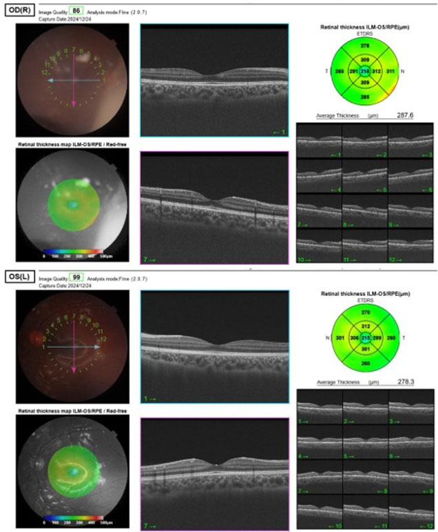
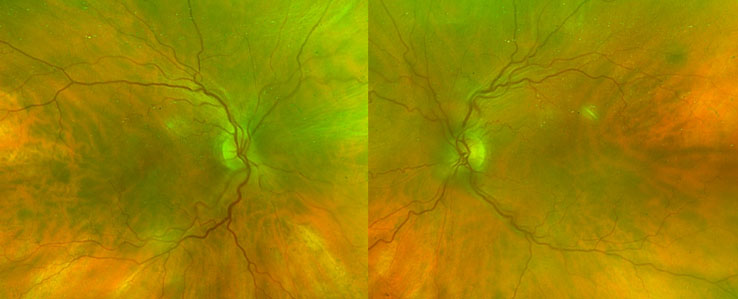
Case Report: A 55-year-old woman underwent tympanoplasty under general anesthesia. Following extubation, the patient reported acute unilateral visual loss immediately after gaining consciousness. Ophthalmic examination revealed a dark coin-shaped premacular hemorrhage resembling sub-ILM hemorrhage in the eye ipsilateral to the operated ear. Findings of multimodal imaging, including optical coherence tomography and fundus autofluorescence, confirmed the diagnosis. The patient was observed without intervention and experienced complete resolution of visual symptoms, with no residual hemorrhage at 3-month follow-up.
Conclusion: We report a case of unilateral sub-ILM hemorrhage occurring shortly after tympanoplasty. Although no established mechanism could be identified, the temporal relationship and ipsilateral presentation raise the possibility of a localized perioperative contribution. To our knowledge, similar cases have not been reported previously. Ophthalmic evaluation should be considered in patients who develop visual symptoms following otologic surgery.
Keywords: Optical coherence tomography, Postoperative visual loss, Sub-internal limiting membrane hemorrhage, Tympanoplasty
SUPPORTING INFORMATION
Author Contributions
Farah AlNasser - Substantial contributions to conception and design, Acquisition of data, Analysis of data, Interpretation of data, Drafting the article, Revising it critically for important intellectual content, Final approval of the version to be published
Lulwa AlMatouq - Substantial contributions to conception and design, Acquisition of data, Analysis of data, Interpretation of data, Drafting the article, Revising it critically for important intellectual content, Final approval of the version to be published
Aseel AlKandari - Substantial contributions to conception and design, Acquisition of data, Analysis of data, Interpretation of data, Drafting the article, Revising it critically for important intellectual content, Final approval of the version to be published
Alaa AlAli - Substantial contributions to conception and design, Acquisition of data, Analysis of data, Interpretation of data, Drafting the article, Revising it critically for important intellectual content, Final approval of the version to be published
Guaranter of SubmissionThe corresponding author is the guarantor of submission.
Source of SupportNone
Consent StatementWritten informed consent was obtained from the patient for publication of this article.
Data AvailabilityAll relevant data are within the paper and its Supporting Information files.
Conflict of InterestAuthors declare no conflict of interest.
Copyright© 2026 Farah AlNasser et al. This article is distributed under the terms of Creative Commons Attribution License which permits unrestricted use, distribution and reproduction in any medium provided the original author(s) and original publisher are properly credited. Please see the copyright policy on the journal website for more information.