|
| Case Report | ||||||
| Bilateral abducens nerve palsy with papilledema following electrical injury: A case report | ||||||
| Yunita Mansyur1, Meiliaty A. Angky1, Batari T. Umar1 | ||||||
| 1Department of Ophthalmology, Faculty of Medicine, Hasanuddin University, Celebes Eye Center, Makassa, Indonesia | ||||||
| ||||||
| [HTML Abstract]
[PDF Full Text][Print This Article] [Similar articles in PubMed][Similar articles in Google Scholar] |
| How to cite this article |
| Mansyur Y, Angky MA, Umar BT. Bilateral abducens nerve palsy with papilledema following electrical injury: A case report. J Case Rep Images Opthalmol 2018;1:100004Z17YM2018 |
| ABSTRACT | ||||||
|
Introduction: Abducens nerve is the cranial nerve most vulnerable to injury. Abducens nerve palsy were commonly found after head injury - about 1% to 2.7% of all head injuries but never been reported caused by electrical injury. This article reports a case of bilateral abducens nerve palsy with papilledema following electrical injury of a 63-year-old woman came with a history of electrical injury two weeks before. There was a history of unconsciousness for three days after injury with no evident of head trauma. Patient complaint double vision after gaining consciousness. Best-corrected visual acuities were 20/20 and 20/25. Eye examination revealed bilateral abducens nerve palsy and funduscopy showed papilledema in both eyes. Head Computed tomography (CT) scan taken one day after injury showed hematoma and cerebral contusion in the bilateral frontal area with subarachnoid hematoma. At six-month follow-up with conservative management, diplopia was fully recovered. Conclusion: This case report highlights the occurrence of bilateral abducens nerve palsy following an electrical injury and conservative management showed good result in this case. Keywords: Bilateral abducens nerve palsy, Electrical injury, Intracranial pressure, Papilledema | ||||||
| INTRODUCTION | ||||||
|
The abducens nucleus is the cranial nerve most vulnerable to injury. Dysfunction of the abducens nerve can result from lesions occurring anywhere along its course between the nucleus in the dorsal pons and the lateral rectus muscle within the orbit [1],[2]. It has been reported that 1% to 2.7% of all head injuries are followed by unilateral abducens palsy, but bilateral abducens nerve palsy is extremely rare [3],[4] . Electrical injury may cause various ocular complications without major damage to other organs of the body. Ocular complications from electrical burn injuries are not very common and have rarely been described. Some ocular manifestations of electrical injury have been reported such as anisocoria, bilateral cataract formation, iritis, and macular cyst formation [5]. Bilateral abducens palsy following an electrical injury has never been reported before. We report a case of bilateral abducens nerve palsy associated with papilledema in a 63-year-old woman presenting with double vision following an electrical injury. | ||||||
| CASE REPORT | ||||||
|
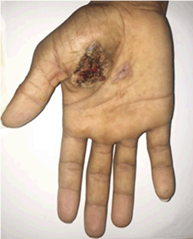
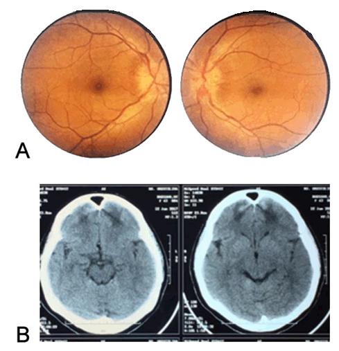
A 63-year-old woman came with a history of electrical injury two weeks before. Patient was injured by electrical shock from electronic devices with 350 watts output. History of unconsciousness for three days. Patient complaint decreased of visual acuity and double vision after gaining consciousness. There was history of vomiting and methemoglobinuria. General physical examination showed healing wound in right hand which was about 5 cm × 4 cm in size (Figure 1). There was no evidence of head trauma. Eye examination revealed best corrected visual acuity on right eye 20/20 and 20/25 on left eye. Diplopia was mostly at distance but was occasionally noted at near. Separation of objects was horizontal. There were bilateral esodeviation with limitation in abduction corresponding to bilateral abducens nerve palsy (Figure 2). The rest of cranial nerves were normally functioned. Pupils were normal. No relative afferent pupillary defect was found. Funduscopy showed papilledema in both eyes (Figure 3A). Head Computed Tomography (CT) scans taken one day after injury showed hematoma and cerebral contusion in the bilateral frontal area and also subarachnoid hematoma. No data except expertation from the radiologist. Second head CT-scan was taken after two weeks after injury that showed resolution of hematoma with focal edema remain in frontal area (Figure 3B). Based on history and clinical examination, provisional diagnosis of bilateral abducens palsy and papilledema was made. No treatment was given for this patient. We planned 3–6 months observation to evaluate spontaneous resolution of bilateral abducens palsy. At 6-month follow-up, diplopia was fully recovered. | ||||||
| ||||||
|
| ||||||
|
| ||||||
DISCUSSION | ||||||
|
Cranial nerve VI, also known as the abducens nerve, innervates the ipsilateral lateral rectus (LR), which functions to abduct the ipsilateral eye. Abducens nerve nucleus is located in the pons, just ventral to the floor of the fourth ventricle and just lateral to the medial longitudinal fasciculus (MLF). About 40% of its neurons project into the ipsilateral MLF only to cross over to the contralateral side and ascend to innervate that contralateral medial rectus subnucleus to participate in contralateral eye adduction [1],[2]. The abducens nerve emerges from the brainstem at the pontomedullary junction to enter the subarachnoid space, coursing upward between the pons and clivus to enter the Dorello canal. This nerve has the longest subarachnoid course of all cranial nerves. At the petrous apex, it angulates to enter the cavernous sinus and travels in close proximity to the internal carotid artery. The abducens nerve then proceeds through the superior orbital fissure and innervates the lateral rectus muscle [1],[2]. Dysfunction of the abducens nerve can result from lesions occurring anywhere along its course between the nerve nucleus in the dorsal pons and the lateral rectus muscle within the orbit [6]. Patients with sixth nerve palsy present with binocular horizontal diplopia, worse in the distance, and esotropia in primary gaze. Examination for sixth nerve palsy involves documenting the presence or absence of papilledema, examining the ocular motility, evaluating the eyelids and pupils, and excluding involvement of other cranial nerves (eg, V, VII, VIII) [2],[6]. In this patient we found bilateral esodeviation with limitation in abduction corresponding to bilateral abducens nerve palsy and also bilateral papiledema but we couldn’t find any abnormal head posture in patient. The rest of cranial nerves were normally functioned. Bilateral palsy might occur as a result of direct injury to both nerves independently or indirectly from changing intracranial pressure [2],[7]. Changes in intracranial pressure either increased or decreased may result in downward displacement of the brainstem causing stretching of the abducens nerve which is tethered as its exits the pons and inside Dorello’s canal [8]. Papilledema in funduscopy became a proof that intracranial pressure increased in this patient which is caused by either the hematoma and cerebral contusion in bilateral frontal area or the subarachnoid hematoma or even both. Nathan et al. postulated several mechanisms to explain the bilateral abducens nerve involvement in subarachnoid hematoma. These included direct compression by the aneurysm, increased intracranial pressure, vasospasm of the pontine branches of the basilar artery affecting the abducens nuclei, and direct compression of the clot on the nerve in the prepontine cistern [9]. We suggested that the sixth nerve palsy were due to increasing of intracranial pressure caused by bleeding in intracranial structures. The bleeding process could be induced by electrical injury through some mechanisms. Electrical injury results in direct damage of the tissue because of the electric current and subsequent heat generation [10]. Heat generation due to the passing current causes direct damage to the tissues. Electrical injuries often result in extensive tissue damage where vascular damage to different layers of the vessel may occur and result in thrombosis and spontaneous rupture of blood vessels [11] . Wang et al. examined aorta and pulmonary artery endothelial cells from electrocution victims and found that cell membrane perforations were present within 24 hours of the electrical injury, whereas after 24 hours, the endothelium disintegrated [11],[12]. Jaffe et al. found that electrical injury caused a complete loss of endothelial cells. The muscle fibers of the media were more sensitive to electric current, whereas the adventitia showed little change. The vessels lost their elasticity, and fusiform aneurysms were common [11],[13]. Theelectrical current that passed through different tissue resistance damaged the vascular tissue. Damaged vessels role as a bleeding source in this case. Treatment of abducens palsy includes prism control, strabismus surgery, and chemodenervation. Although the overall spontaneous recovery rate of traumatic abducens nerve palsy is high, a complete or bilateral case has a poor recovery rate [14], [15], [16]. Mutyala et al reported 72% spontaneous resolution in unilateral cases against only 12% in bilateral cases (6-month follow-up period) [17]. In our case, we planned to observe spontaneous resolution in six months period. At 6-month follow-up, diplopia was fully recovered. In this case, bilateral palsy recovered since it were incomplete palsy and the cause of increased intracranial pressure started to resolve resulting in good prognosis. | ||||||
| CONCLUSION | ||||||
|
Bilateral abducens nerve palsy may happen after an electrical injury and is an extreme rare case. The proposed mechanism of bilateral nerve palsy in electrical injury was through the damaged of brain vascular from electrical current, caused bleeding in intracranial structures which then increased the intracranial pressure and stretched the bilateral abducens nerve. Our patient was managed conservatively and diplopia was fully recovered at 6-month follow-up. This case report highlights the occurrence and management of bilateral abducens nerve palsy following an electrical injury. | ||||||
| REFERENCES | ||||||
| ||||||
|
[HTML Abstract]
[PDF Full Text]
|
| Author Contributions
Yunita Mansyur – Substantial contributions to conception and design, Acquisition of data, Analysis and interpretation of data, Drafting the article, Revising it critically for important intellectual content, Final approval of the version to be published Meiliaty A. Angky – Substantial contributions to conception and design, Acquisition of data, Analysis and interpretation of data, Drafting the article, Revising it critically for important intellectual content, Final approval of the version to be published Batari T. Umar – Substantial contributions to conception and design, Acquisition of data, Analysis and interpretation of data, Drafting the article, Revising it critically for important intellectual content, Final approval of the version to be published |
|
Guarantor of Submission
The corresponding author is the guarantor of submission. |
|
Source of Support
None |
|
Consent Statement
Written informed consent was obtained from the patient for publication of this case report. |
|
Conflict of Interest
Author declares no conflict of interest. |
|
Copyright
© 2018 Yunita Mansyur et al. This article is distributed under the terms of Creative Commons Attribution License which permits unrestricted use, distribution and reproduction in any medium provided the original author(s) and original publisher are properly credited. Please see the copyright policy on the journal website for more information. |
|
|